

When to Order Imaging for Acute Right Upper Quadrant Abdominal Pain
Whether you are in the emergency department, hospital, or an outpatient setting, right upper quadrant pain is a common concern. When should we order diagnostic imaging and what test is the right test to order?
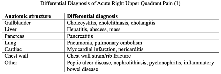
Case: You’re seeing a 50-year-old male with acute right upper quadrant pain. It started 24 hours ago, with nausea and vomiting. What is your initial differential diagnosis? Let’s break it down by the anatomic framework. What organs/structures are in the right upper quadrant of the abdomen?
Back to the case: from your history and physical exam, pertinent positive findings include a heart rate of 110, temperature of 38.5C (101.3F), right upper quadrant pain on palpation with guarding and positive murphy’s sign (deep palpation of the right upper quadrant followed by the examiner asking the patient to inspire - if the patient’s breath abruptly stops, it is a positive finding). Pertinent negatives include no shortness of breath, cough, leg swelling, no chest wall pain on palpation, no lower urinary tract symptoms, diarrhea, melena, or bright red blood per rectum. He does not appear to be jaundiced. The remainder of the physical exam is unremarkable. The primary diagnostic concern is acute cholecystitis.
What is acute cholecystitis? It is inflammation and infection of the gallbladder that occurs when gallstones and/or biliary sludge is blocking the cystic duct or impairing emptying of the gallbladder (2).
Diagnostic Approach to cholecystitis: many patients with acute cholecystitis will present with right upper quadrant pain, nausea, vomiting, appetite loss and fever (3). The diagnosis of acute cholecystitis cannot be made on history and physical examination alone, and imaging is recommended as part of the work up (3, 4).
Role of Imaging (in adults, excluding pregnant patients): If we suspect biliary disease, the initial imaging modality of choice is the abdominal ultrasound - it is highly accurate at diagnosing or excluding gallstones, and may differentiate cholelithiasis from gallbladder sludge, polyps, or masses (2). When performing the ultrasound, the sonographic murphy’s sign is also elicited (the sonographic equivalent to the clinical murphy’s sign, where the ultrasound probe is used instead of the examiners hand). Abdominal ultrasound has excellent test characteristics to rule in or out cholecystitis (5). It is also the least expensive and usually the quickest imaging modality.
Is there a role for CT? If the abdominal ultrasound is negative or equivocal, what do we do next? CT scans will have inferior diagnostic accuracy for cholecystitis, however, will identify other potential pathology causing the right upper quadrant pain so should be considered (3). Non-contrast CT adds limited value to assess for biliary sources of right upper quadrant pain (3).
Is there a role for MRI? A noncontrast MRI abdmen with MR cholangiopancreatography (MRCP) has excellent accuracy for detecting biliary stones (this test does not use IV contrast). It is especially useful to evaluate stones in the cystic duct and common bile duct (3).
Are there other modalities to assess for acute cholecystitis? Yes. Let’s summarize with a table.

Back to the case: you are able to get a rapid abdominal ultrasound, which reveals the following:
Gallbladder wall thickening
Stones present in the gallbladder
Pericholecystic fluid
Sonographic Murphy’s sign
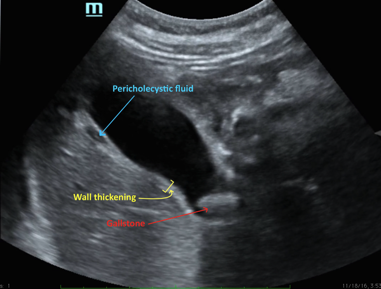
Source: Correa & Spiegleman
This is a slam dunk case of acute cholecystitis, and a referral to general surgery for management is warranted.
Key Take Home Point: if we suspect acute cholecystitis, the abdominal ultrasound is the initial diagnostic imaging choice, and is a necessary part of the diagnostic pathway.
If you were wondering about lab tests for the evaluation of acute right upper quadrant abdominal pain, this week’s NP Reasoning newsletter will review pertinent labs and interpretation tips. Sign up today for free!
